Abstract
The state of deprotonation/protonation of surfaces has far-ranging implications in chemistry, from acid–base catalysis1 and the electrocatalytic and photocatalytic splitting of water2, to the behaviour of minerals3 and biochemistry4. An entity’s acidity is described by its proton affinity and its acid dissociation constant pKa (the negative logarithm of the equilibrium constant of the proton transfer reaction in solution). The acidity of individual sites is difficult to assess for solids, compared with molecules. For mineral surfaces, the acidity is estimated by semi-empirical concepts, such as bond-order valence sums5, and increasingly modelled with first-principles molecular dynamics simulations6,7. At present, such predictions cannot be tested—experimental measures, such as the point of zero charge8, integrate over the whole surface or, in some cases, individual crystal facets9. Here we assess the acidity of individual hydroxyl groups on In2O3(111)—a model oxide with four different types of surface oxygen atom. We probe the strength of their hydrogen bonds with the tip of a non-contact atomic force microscope and find quantitative agreement with density functional theory calculations. By relating the results to known proton affinities of gas-phase molecules, we determine the proton affinity of the different surface sites of In2O3 with atomic precision. Measurements on hydroxylated titanium dioxide and zirconium oxide extend our method to other oxides.
Access options
Subscribe to Journal
Get full journal access for 1 year
199,00 €
only 3,90 € per issue
Tax calculation will be finalised during checkout.
Rent or Buy article
Get time limited or full article access on ReadCube.
from$8.99
All prices are NET prices.


Data availability
The datasets generated and analysed during the current study are available from the corresponding author on reasonable request.
References
- 1.
Kramer, G. J., van Santen, R. A., Emeis, C. A. & Nowak, A. K. Understanding the acid behaviour of zeolites from theory and experiment. Nature 363, 529–531 (1993).
- 2.
She, Z. W. et al. Combining theory and experiment in electrocatalysis: insights into materials design. Science 355, eaad4998 (2017).
- 3.
Brown, G. E. Jr & Calas, G. Mineral–aqueous solution interfaces and their impact on the environment. Geochem. Perspect. 1, 483–742 (2012).
- 4.
Silverman, R. B. Organic Chemistry of Enzyme-Catalyzed Reactions (Academic Press, 2002).
- 5.
Hiemstra, T., Venema, P. & Riemsdijk, W. Intrinsic proton affinity of reactive surface groups of metal (hydr)oxides: the bond valence principle. J. Colloid Interface Sci. 184, 680–692 (1996).
- 6.
Cheng, J. & Sprik, M. Acidity of the aqueous rutile TiO2(110) surface from density functional theory based molecular dynamics. J. Chem. Theory Comput. 6, 880–889 (2010).
- 7.
Gittus, O. R. von Rudorff, G. F., Rosso, K. M. & Blumberger, J. Acidity constants of the hematite–liquid water interface from ab initio molecular dynamics. J. Phys. Chem. Lett. 9, 5574–5582 (2018).
- 8.
Kosmulski, M. Compilation of PZC and IEP of sparingly soluble metal oxides and hydroxides from literature. Adv. Colloid Interface Sci. 152, 14–25 (2009).
- 9.
Bullard, J. W. & Cima, M. J. Orientation dependence of the isoelectric point of TiO2 (rutile) surfaces. Langmuir 22, 10264–10271 (2006).
- 10.
Giessibl, F. J. The qPlus sensor, a powerful core for the atomic force microscope. Rev. Sci. Instrum. 90, 011101 (2019).
- 11.
Gross, L., Mohn, F., Moll, N., Liljeroth, P. & Meyer, G. The chemical structure of a molecule resolved by atomic force microscopy. Science 325, 1110–1114 (2009).
- 12.
Setvín, M. et al. Polarity compensation mechanisms on the perovskite surface KTaO3(001). Science 359, 572–575 (2018).
- 13.
Peng, J. et al. The effect of hydration number on the interfacial transport of sodium ions. Nature 557, 701–705 (2018); correction 563, E18 (2018).
- 14.
Lantz, M. A. et al. Quantitative measurement of short-range chemical bonding forces. Science 291, 2580–2583 (2001).
- 15.
Sugimoto, Y. et al. Chemical identification of individual surface atoms by atomic force microscopy. Nature 446, 64–67 (2007).
- 16.
Gross, L. et al. Measuring the charge state of an adatom with noncontact atomic force microscopy. Science 324, 1428–1431 (2009).
- 17.
Onoda, J., Ondráček, M., Jelínek, P. & Sugimoto, Y. Electronegativity determination of individual surface atoms by atomic force microscopy. Nat. Commun. 8, 15155 (2017).
- 18.
Hunter, E. P. L. & Lias, S. G. Evaluated gas phase basicities and proton affinities of molecules: an update. J. Phys. Chem. Ref. Data 27, 413–656 (1998).
- 19.
Gorte, R. J. & White, D. Interactions of chemical species with acid sites in zeolites. Top. Catal. 4, 57–69 (1997).
- 20.
Larrazábal, G. O., Shinagawa, T., Martín, A. J. & Pérez-Ramírez, J. Microfabricated electrodes unravel the role of interfaces in multicomponent copper-based CO2 reduction catalysts. Nat. Commun 9, 1477 (2018).
- 21.
Martin, O. et al. Indium oxide as a superior catalyst for methanol synthesis by CO2 hydrogenation. Angew. Chem. Int. Ed. 128, 6369–6373 (2016).
- 22.
Hagleitner, D. R. et al. Bulk and surface characterization of In2O3(001) single crystals. Phys. Rev. B 85, 115441 (2012).
- 23.
Wagner, M. et al. Reducing the In2O3(111) surface results in ordered indium adatoms. Adv. Mater. Interfaces 1, 1400289 (2014).
- 24.
Capdevila-Cortada, M., Vilé, G., Teschner, D., Pérez-Ramírez, J. & López, N. Reactivity descriptors for ceria in catalysis. Appl. Catal. B 197, 299–312 (2016).
- 25.
Wagner, M. et al. Resolving the structure of a well-ordered hydroxyl overlayer on In2O3(111): nanomanipulation and theory. ACS Nano 11, 11531–11541 (2017).
- 26.
Yurtsever, A. et al. Understanding image contrast formation in TiO2 with force spectroscopy. Phys. Rev. B 85, 125416 (2012).
- 27.
Stetsovych, O. et al. Atomic species identification at the (101) anatase surface by simultaneous scanning tunnelling and atomic force microscopy. Nat. Commun. 6, 7265 (2015).
- 28.
Sugimoto, Y. et al. Quantum degeneracy in atomic point contacts revealed by chemical force and conductance. Phys. Rev. Lett. 111, 106803–106805 (2013).
- 29.
Kowalski, P. M., Meyer, B. & Marx, D. Composition, structure, and stability of the rutile TiO2(110) surface: oxygen depletion, hydroxylation, hydrogen migration, and water adsorption. Phys. Rev. B 79, 115410 (2009).
- 30.
Lackner, P. et al. Water adsorption at zirconia: from the ZrO2(111)/Pt3Zr(0001) model system to powder samples. J. Mater. Chem. A 6, 17587–17601 (2018).
- 31.
Huber, F. & Giessibl, F. J. Low noise current preamplifier for qPlus sensor deflection signal detection in atomic force microscopy at room and low temperatures. Rev. Sci. Instrum. 88, 073702 (2017).
- 32.
Štubian, M., Bobek, J., Setvín, M., Diebold, U. & Schmid, M. Fast low-noise transimpedance amplifier for scanning tunneling microscopy and beyond. Rev. Sci. Instrum. 91, 074701 (2020).
- 33.
Sader, J. E. & Jarvis, S. P. Accurate formulas for interaction force and energy in frequency modulation force spectroscopy. Appl. Phys. Lett. 84, 1801–1803 (2004).
- 34.
Giannozzi, P. et al. Quantum ESPRESSO: a modular and open-source software project for quantum simulations of materials. J. Phys. Condens. Matter 21, 395502 (2009).
- 35.
Perdew, J., Burke, K. & Ernzerhof, M. Generalized gradient approximation made simple. Phys. Rev. Lett. 77, 3865–3868 (1996).
- 36.
Vanderbilt, D. Soft self-consistent pseudopotentials in a generalized eigenvalue formalism. Phys. Rev. B 41, 7892–7895 (1990).
- 37.
Wagner, M. et al. Well-ordered In adatoms at the In2O3(111) surface created by Fe deposition. Phys. Rev. Lett. 117, 206101 (2016).
- 38.
Grimme, S., Antony, J., Ehrlich, S. & Krieg, H. A consistent and accurate ab initio parametrization of density functional dispersion correction (DFT-D) for the 94 elements H–Pu. J. Chem. Phys. 132, 154104 (2010).
- 39.
Sokolović, I. et al. Resolving the adsorption of molecular O2 on the rutile TiO2(110) surface by noncontact atomic force microscopy. Proc. Natl Acad. Sci. USA 117, 14827–14837 (2020).
- 40.
Hapala, P. et al. Mechanism of high-resolution STM/AFM imaging with functionalized tips. Phys. Rev. B 90, 085421 (2014).
- 41.
Linstrom, P. J. & Mallard, W. G. (eds) NIST Chemistry WebBook: NIST Standard Reference Database Number 69 (National Institute of Standards and Technology, 2018); https://webbook.nist.gov/chemistry.
Acknowledgements
This work was supported by the Austrian Science Fund (FWF), project V 773-N (Elise-Richter-Stelle, M.W.) and Z 250-N27 (Wittgenstein Prize, U.D.), as well as the German Research Foundation (DFG), Research Unit FOR 1878 (funCOS, B.M.). M.W. and U.D. also acknowledge funding under the Horizon 2020 Research and Innovation Programme under the grant agreement number 810626. M. Setvin acknowledges the support of GAUK Primus/20/SCI/009. Computational resources were provided by LRZ Garching (project pn98fa)and RRZ Erlangen.
Author information
Affiliations
Contributions
M.W. and M. Setvin conducted the experiments. M.W., M. Setvin and M. Schmid analysed the AFM data. B.M. performed the calculations and derived the scaling relationships. M.W., B.M. and U.D. wrote the manuscript, which was reviewed and edited by all authors. U.D. oversaw the project.
Corresponding author
Ethics declarations
Competing interests
The authors declare no competing interests.
Additional information
Peer review information Nature thanks Leo Gross and the other, anonymous, reviewer(s) for their contribution to the peer review of this work.
Publisher’s note Springer Nature remains neutral with regard to jurisdictional claims in published maps and institutional affiliations.
Extended data figures and tables
Extended Data Fig. 1 The clean In2O3(111) surface imaged with various AFM tip terminations.
a, b, Structural models of the In2O3(111) surface, with emphasis on the surface O (red) atoms (a) and In (blue, green) atoms (b). Also indicated is the unit cell. c, AFM image with an O-terminated tip. The intentional functionalization was performed on a reduced rutile TiO2(110) surface with adsorbed O2 molecules by applying voltage pulses (approximately +3 V) above the molecular oxygen species. Previous experience has shown that such a tip functionalization provides excellent resolution of the oxygen sublattice in the repulsive regime39. Such tips provide negligible attractive interaction with the anion lattice and they are rigid; the resulting images are therefore not distorted by bending the tip apex40 and clearly show the O sublattice, O(α) to O(δ). d, AFM image taken after the tip was gently pushed into the hydroxylated In2O3(111) surface to induce an OH termination. The more flexible tip termination leads to crests in the images. The dark, round features correspond to In, which are indicated by the green and blue dots (attractive interaction between negatively charged (tip) OH and In anions). The bright maxima correspond to O(3c).
Extended Data Fig. 2 Distance-dependent STM/AFM images of the clean In2O3(111) surface.
a, b, O-terminated tip. a, Constant-height STM image (tunnelling current, IT). b, Constant-height AFM images (frequency shift, Δf). From top to bottom, the tip–sample distance was progressively reduced in steps of about 50 pm. c, d, OH-terminated tip. c, Constant-current STM image. d, Same as c but the distance was reduced in steps of about 40 pm.
Extended Data Fig. 3 Adsorption sites of OH groups on In2O3(111).
a–f, Constant-height nc-AFM images of three dissociated water molecules in adjoining unit cells, taken with decreasing tip height. The height difference ∆z between the subsequent images is 10 to 15 pm. Each dissociated water molecule gives rise to two OH groups adsorbed next to each other, OWH and OSH (where the ‘W’ and ‘S’ indicate the origin of the oxygen atom, that is, the water molecule or the surface, respectively)25. In a, with the tip farthest away, both OH groups are imaged as dark features. As the tip comes closer to the OH groups (b, c), the OWH (sticking farther away from the surface than the OSH) turn bright (onset of repulsive interaction, but overall attractive forces). In d, the OSH also start to be imaged as bright features. Approaching the surface even more, e, f reveal the O(3c) lattice atoms of the surface; this information was used to determine the adsorption sites of the OH groups experimentally. g, Profiles of the frequency shift across the OSH and OWH pair as indicated by the arrows in a–f. h, Same image as f with the O(3c) lattice superimposed (O(α), white; O(β), red; O(γ), orange; O(δ), yellow). The site of the OSH is identified as an O(β) (circle filled black). i, Atomic model of the surface including the adsorption sites of the OSH and OWH (black circle, filled white), which bridges two In(5c) atoms nearby. The adsorption sites agree with previous STM and DFT results in ref. 25.
Extended Data Fig. 4 The hydroxylated In2O3(111) surface with increasing water coverages in STM and AFM.
The STM (top) and AFM images (bottom) were acquired at different regions of the surface. a1, Oxygen-terminated tip. b1–d1, OH-terminated tips. a, a1, Clean In2O3(111) surface. The contrast in STM (empty states) is dominated by the high density of states of the In(5c) and the lower density of states at the In(6c), which gives dark triangles. In AFM, the contrast is dominated by the topmost atoms of the surface, that is, the 12 O(3c) per unit cell. b, b1, Single dissociated water molecules, OWH and OSH. c, c1, Two and three dissociated water molecules per unit cell. d, d1, Saturation with three dissociated water molecules per unit cell in symmetry-equivalent sites, giving rise to a ‘propeller-like feature’ consisting of three brighter (OWH) and darker (OSH at O(β) site) at equivalent positions. For DFT calculations and structural relaxations see ref. 25.
Extended Data Fig. 5 Manipulation of OSH groups by voltage pulses.
a, STM image before (a1) and during (a2) the manipulation. Five pulses (two times +2.8 V and three times +2.7 V, 20 pA, marked with crosses) were applied during STM imaging in the centre of individual propeller-like structures. b, c, AFM image of the same surface area before the manipulation (b; with crosses marking the position of the pulses) and after the manipulation (c). d, e, Cartoons identifying the various species before (d) and after (e) the manipulation. In each manipulation, one H per propeller was removed, leaving behind a denuded O(β), indicated in e and visible as a smaller, bright dot in the AFM image in c. Two H have desorbed and three H have re-adsorbed, visible as very dark features in AFM in c and indicated in yellow in e.
Extended Data Fig. 6 Data evaluation of F(z) curves.
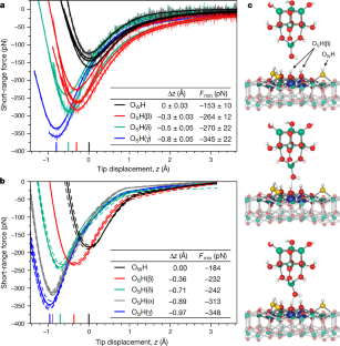
a, Frequency-shift curves acquired on different OH groups and the In2O3(111) background measured in-between the OH groups (mostly in regions A and C, see Fig. 1). b, The averaged curves from a (solid lines) and after background subtraction (dashed lines). c, Background-corrected F(z) curves obtained from b using Sader’s formula33 (fR = 77.7 kHz, k = 5,400 N m−1, A = 100 pm). d, The F(z) curves of Fig. 2a showing the whole z range.
Extended Data Fig. 7 Transferability and reproducibility of the method.
F(z) curves on OSH/TiO2(110) and OH/zirconium oxide/Pt3Zr(0001) obtained with the OH-terminated indium oxide tip. a, TiO2(110): experimental data. The tip was prepared on hydroxylated In2O3(111), and F(z) curves were taken on the OWH and OSH(β) (labelled as such) to ascertain the tip termination (fR = 69.6 kHz, k = 5,400 N m−1, A = 60 pm). For reference, the curves from the main text (Fig. 2) are also plotted on the left; for better visibility they are shifted horizontally by −2 Å. This tip was used to obtain the F(z) curve on OSH/TiO2(110) (brown, shifted to the right). Approaching the OSH on TiO2 even closer results in picking up the H. The hydroxylated TiO2(110) surface was prepared following a well established procedure26, which results in one type of hydroxyl at a bridging O(2c) atom. b, Calculated short-range F(z) curves on bridging hydroxyls on TiO2(110) for two OH coverages (1/4 and 1/8 monolayer) obtained with the OH-terminated InOx tip. c, Zirconium oxide: the standard tip was prepared on hydroxylated In2O3(111), and F(z) curves were taken on the OWH and OSH(β) (labelled as such) to ascertain the tip termination. For reference, the curves from the main text (Fig. 2) are also plotted on the left; for better visibility they are shifted horizontally by −2 Å. This tip was used to take F(z) curves on hydroxylated zirconium oxide (purple, shifted to the right). d, Zirconium oxide: the same type of measurement, but with an unknown tip termination that gives more shallow minima for OWH and OSH on hydroxylated In2O3(111). Note that both tips measure the same relative positions in the force minima of the strongly bound H on In2O3(111) and zirconium oxide, that is, the force on OH/zirconium oxide lies between OWH and OSH(β) (fR = 77.2 kHz, k = 5,400 N m−1, A = 240 pm (In2O3), 250 pm (zirconium oxide)). The ultrathin zirconium oxide layer was prepared following the method of ref. 30. The surface was exposed to 2 langmuir (1 L = 1.33 × 10⁻⁶ mbar s) of water at 320 K.
Extended Data Fig. 8 Reactivity of the hydroxylated surface.
a, Atom-resolved PDOS of the four symmetry-inequivalent three-fold coordinated O surface sites on the fully hydroxylated In2O3(111) surface with three OWH and three OSH(β) per unit cell. The VBM is at 0 eV. b, Calculated adsorption energies \({E}_}^}\) for the remaining three unprotonated O(3c) sites (with respect to the H2 gas phase molecule). The adsorption of water and the formation of the hydroxylated surface structure slightly modifies the reactivity of the unprotonated O(α), O(γ) and O(δ) sites. The saturation of the O(β) sites by protons leads to a strong downward shift of the O(β) 2p states. The VBM is now formed by the O(δ) 2p states, followed by the O 2p states from the O(α) and O(γ) sites. The O(δ), which are the second-most-reactive O species on the uncovered surface, are thus expected to be the most reactive sites on the hydroxylated In2O3(111) surface. This is confirmed by the calculated H adsorption energies (b). Whereas for O(γ) the H adsorption energies are rather similar on the clean and on the hydroxylated surface, the local relaxations upon water adsorption make the O(α) sites slightly more and the O(δ) slightly less reactive (see b and inset in Fig. 1b). As for the clean surface, the pronounced peak in the PDOS of the three-fold coordinated OS surface atoms at the VBM also leads to an upward shift of their p-band centre. The shift of the p-band centre has been taken as an indirect measure of surface reactivity24 and confirms the expected trend in the PA of the different surface sites.
Extended Data Fig. 9 Tip model for the AFM calculations.
a, Relaxed structure of an In8O12 cluster cut out of an In2O3(111) slab. The cut is centred around the high-symmetry B site of the surface and its depth is four In2O3(111) trilayers. The In2O3(111) trilayers are all equivalent but they are shifted such that the high-symmetry sites with a three-fold symmetry axis (see Fig. 1a) form a stacking sequence of BCAB. The cluster consists of an In atom at the B site together with three O atoms, and six-membered In3O3 rings around the A and C sites. The cluster is stoichiometric and stable upon geometry optimization, it has a three-fold symmetry axis and its HOMO–LUMO gap is 1.62 eV (PBE). b, Cluster after dissociative adsorption of six water molecules to saturate the broken In–O bonds. The OH groups were added to the six In atoms in the two central planes, and the protons were placed on top of the two-fold bridging O atoms. This increased the HOMO–LUMO gap to 2.57 eV. In the next step, the upper In(OH)3 cap was removed (dashed circle). This was done to eliminate the top three-fold coordinated In atom and to reduce the height of the tip model. This saves some computer time in the AFM calculations, as the thickness of the vacuum region can be reduced. Removing the cap increases the HOMO–LUMO gap to 2.68 eV. At this point, the cluster still has a three-fold symmetry axis. c, Structure of the final tip. One additional water molecule is added to the cluster (b). The OH group is adsorbed at the In apex atom of the tip and the proton is added to one of the remaining O atoms. This breaks the three-fold symmetry of the tip. Careful test calculations showed that the specific choice for the adsorption site of the proton is not important. All tips gave basically the same F(z) curves. In the end, the proton was split into three parts, and three pseudo-hydrogen atoms with nuclear charge of +1/3 were added to the three O atoms closest to the tip apex indicated by arrows. The HOMO–LUMO gap of the final cluster calculated with the PBE functional is 2.64 eV.
Extended Data Fig. 10 Illustration of the background subtraction for the calculated F(z) curves.
Grey dashed lines are calculated F(z) curves at the high-symmetry A and C sites on the hydroxylated and the water-free In2O3(111) surface, each for two different azimuthal tip orientations. The average of the eight curves is shown as a grey solid line. In the background-correction procedure, this curve is subtracted from all calculated F(z) curves above surface OH groups. Black and red graphs show the data for the OWH and OSH(β) groups before background subtraction (dashed, calculated curves for two tip orientations; solid, average of the two dashed curves).
Extended Data Fig. 11 Correlations between force and energy minima, PA and pKa, using probe molecules.
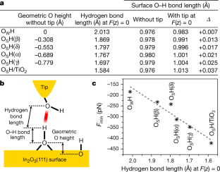
a, List of probe molecules with their experimental PA and pKa values. As our aim is to assign PAs to surface O species, only oxygen-based acids and alcohols were selected. The experimental PAs (for the corresponding base, that is, the X−O− anion) are taken from the National Institute of Standards and Technology (NIST) database41. The number of listed NIST entries is given in parenthesis. In case that data from several experiments are available, averaged values and uncertainties are given. b, Correlation between force and energy minima. The linear fit to the calculated minima of the E(z) and F(z) curves for our probe molecules (red dashed line) shows an almost perfect linear correlation of the data points. The value of the energy minimum in the E(z) curves (that is, F(z) = 0) is the natural measure for the strength of the hydrogen bond that forms between the OH groups of the tip and the molecules. This linear relationship allows us to focus on establishing a correlation between PA or acidity and the force, and not the energy minimum. This is more convenient, because the force minimum is the natural measure from the AFM experiments. Although the energy minimum can be obtained by integrating the F(z) curves, it is not trivial to eliminate ambiguities stemming from a proper choice for the zero point of energy, which determines the integration constant. Furthermore, the force minimum is encountered at a larger OH–tip distance than the energy minimum. The force minimum is therefore less influenced by other interactions between the tip and side groups of the molecule or neighbouring atoms on the surface. c, Correlation between the PA and acidity constant pKa of the selected probe molecules. The PA also governs the pKa in wet, solution-based chemical processes: an OH group with a strongly bound proton (high PA of the O atom) is a weak acid, and an OH group with a weakly bound proton (low PA of the O atom) is a strong acid. A linear fit (red dashed line) to the experimental data in a shows the expected trend, that is, strong acids have a low PA, and weak acids bind their proton more strongly. However, in addition to the PA, the pKa also includes the Gibbs free energy of solvation of the acid (XOH), the conjugated base (XO−) and the H+ (see the thermodynamic cycle in Methods). These species have different solubilities, which leads to a large scatter of the data points. d, Correlation between calculated AFM force minima and the experimental acidity constants pKa of the probe molecules. As expected from the discussion in c, the force minima show a larger scatter around the red dashed regression line than when plotted with respect to the PA (see Fig. 4). This is because the AFM measurements are done in vacuum and do not include information about solvation free energies. Thus, the prediction of absolute pKa values for our OH groups on the In2O3(111) surface based on the AFM measurements alone is not possible. Still, there is a clear trend that strong acids form strong H bonds with the AFM tip (deep force minimum, low PA), whereas weak acids form weak H bonds (shallow force minimum, high PA). If we assume that the solvation energies of the structurally rather similar OH groups do not differ too much, then the deviation from the regression line would be similar for all of them, which would allow us to predict at least relative pKa changes between the OH groups by using the slope of the linear regression. The AFM-measured difference in the force minima of 81 pN for the OSH(β) and OSH(γ) sites (see Fig. 2) then translates to a difference in acidity of 5.5 pKa units, which is reasonable.
Supplementary information
AFM tip approaching an O
Supplementary Video 1 SH(β) hydroxyl. Sequence of configurations from the DFT calculation of a force-distance curve between an OSH(β) surface hydroxyl and an OH-terminated indium oxide tip. When the tip approaches the surface, a hydrogen bond forms between the O atom at the tip apex and the proton of the surface hydroxyl.
AFM tip approaching an O
Supplementary Video 2 WH hydroxyl. Sequence of configurations from the DFT calculation of a force-distance curve between an OWH surface hydroxyl and an OH-terminated indium oxide tip. When the tip approaches the surface, a hydrogen bond forms between the O atom at the tip apex and the proton of the surface hydroxyl.
Rights and permissions
About this article
Cite this article
Wagner, M., Meyer, B., Setvin, M. et al. Direct assessment of the acidity of individual surface hydroxyls. Nature 592, 722–725 (2021). https://ift.tt/3gITGMq
-
Received:
-
Accepted:
-
Published:
-
Issue Date:
Comments
By submitting a comment you agree to abide by our Terms and Community Guidelines. If you find something abusive or that does not comply with our terms or guidelines please flag it as inappropriate.
"direct" - Google News
April 28, 2021 at 10:16PM
https://ift.tt/3nrqivw
Direct assessment of the acidity of individual surface hydroxyls - Nature.com
"direct" - Google News
https://ift.tt/2zVRL3T
https://ift.tt/2VUOqKG
Direct
Bagikan Berita Ini


0 Response to "Direct assessment of the acidity of individual surface hydroxyls - Nature.com"
Post a Comment